📋 Important Reminder
Your MRI report is written by a radiologist for your referring doctor—not directly for patients. That's why it contains medical terminology that can sound confusing or alarming. This guide will help you understand the basics, but always discuss your results with your physician, who knows your full medical history and can put findings in proper context.
📑 Quick Navigation

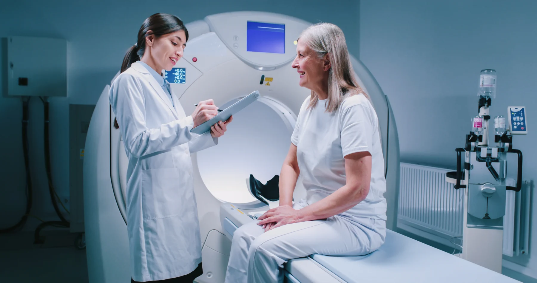
Your doctor will review your MRI results and explain what they mean for your specific situation
You've had your MRI, and now you have a report full of medical jargon like "hyperintense signal" and "mild degenerative changes." It can feel overwhelming—and sometimes scary. But here's the truth: many common findings sound worse than they actually are. Let's break down what radiologists are really saying in plain English.
1 How to Read Your MRI Report
Every MRI report follows a standard format. Here's what each section means:
Clinical History / Indication
Why you got the MRI
Example:
"45-year-old female with chronic lower back pain radiating down left leg. Rule out disc herniation."
What this means: This section tells the radiologist what your doctor is concerned about so they know what to look for. It's based on your symptoms and your doctor's examination.
Technique
Technical details of how the scan was done
Example:
"MRI of the lumbar spine was performed without contrast on a 1.5 Tesla magnet using standard protocols including T1, T2, and STIR sequences."
What this means: This is for medical documentation—you can skip this section! It just describes the MRI machine strength and the types of images taken.
Comparison
Previous imaging (if any)
Example:
"Comparison made with lumbar spine MRI dated 06/15/2023."
What this means: If you've had MRIs before, the radiologist compares them to see if anything has changed. This is actually very helpful—it shows whether findings are new, worse, better, or stable.
Findings
THE MOST IMPORTANT SECTION—What the radiologist sees
Example:
"There is a left paracentral disc herniation at L4-L5 with mild central canal stenosis and left foraminal narrowing. Mild degenerative disc disease is noted at L5-S1. No acute fracture or marrow abnormality."
What this means: This is where the radiologist describes everything they see—both normal and abnormal. It's written in medical terminology, but we'll translate the common terms below! This section goes body part by body part.
Impression / Conclusion
THE SUMMARY—Start here!
Example:
"1. L4-L5 left paracentral disc herniation with mild canal
stenosis and left neural foraminal narrowing, likely
contributing to patient's symptoms.
2. Mild degenerative changes at L5-S1.
3. No acute fracture."
What this means: This is the TL;DR (summary) of the findings. It's numbered by importance, with the most significant findings listed first. Your doctor focuses mainly on this section.
💡 Pro Tip: Start at the Bottom
Most patients find it easier to read the Impression/Conclusion first (the summary at the bottom), then go back and read the detailed Findings section if they want more information. The Impression tells you what matters most.
2 Medical Term Translator: What It Really Means
Here's a plain-English translation of the most common terms you'll see in MRI reports:
📊 Words Describing Severity
Translation: Minor, small, slight. Usually not a big concern. Often age-related wear and tear.
Translation: Medium level. More significant than mild but not severe. May or may not cause symptoms.
Translation: Marked, pronounced. The most significant level. More likely to cause symptoms or require treatment.
Translation: Very small, barely there. Usually insignificant.
🦴 Common Spine Terms
| Medical Term | Plain English |
|---|---|
| Disc herniation / bulge | The cushion between spine bones is pushing out. "Herniation" is more severe than "bulge." |
| Degenerative disc disease | Normal wear-and-tear aging of the spine. Like getting wrinkles—happens to everyone over time. |
| Stenosis | Narrowing of a space. "Spinal stenosis" = the spinal canal is narrower than normal. |
| Foraminal narrowing | The holes where nerves exit the spine are smaller than they should be. Can pinch nerves. |
| Spondylosis | General term for age-related spine arthritis and degeneration. |
| Facet arthropathy | Arthritis in the small joints of the spine. Very common with aging. |
| Spondylolisthesis | One vertebra has slipped forward on the one below it. Can be mild (grade 1) to severe (grade 4). |
| Neural impingement | A nerve is being touched or compressed. This can cause pain, numbness, or tingling. |
🦵 Common Joint Terms (Knee, Shoulder, etc.)
| Medical Term | Plain English |
|---|---|
| Meniscal tear | The cartilage cushion in your knee is torn. Very common, especially with aging or sports. |
| Rotator cuff tear | One or more of the shoulder muscles/tendons is torn. Can be partial or complete (full-thickness). |
| Tendinosis / tendinopathy | The tendon is worn down or irritated. Chronic overuse injury (not acute inflammation). |
| Effusion | Fluid buildup in the joint. "Joint swelling." |
| Chondromalacia | Softening or damage to the cartilage. Common behind the kneecap (patella). |
| Labral tear | The ring of cartilage around the shoulder or hip socket is torn. |
| Osteoarthritis | Wear-and-tear arthritis. Cartilage breakdown and bone changes. Very common with aging. |
🧠 Common Brain Terms
| Medical Term | Plain English |
|---|---|
| White matter changes | Small areas of different signal in the brain's "wiring." Very common with aging and often benign. |
| Nonspecific T2/FLAIR hyperintensities | Bright spots on certain MRI sequences. Usually age-related or from migraines. Often not significant. |
| Microvascular ischemic changes | Tiny areas of reduced blood flow, typically from aging or high blood pressure. Very common in older adults. |
| Cerebral atrophy | Brain shrinkage. Normal part of aging, though can be accelerated in some conditions. |
| Sinusitis | Inflammation or fluid in the sinus cavities. Often an incidental finding. |
📡 MRI Signal Terms
Hyperintense (or "increased signal")
Appears bright/white on the MRI image. What this means depends on the sequence and body part.
Hypointense (or "decreased signal")
Appears dark/black on the MRI image. Again, meaning varies by context.
Enhancement
An area that lights up after contrast dye is given. Can indicate inflammation, infection, or tumor (radiologist will specify).
Don't worry too much about signal terms. Radiologists use these to describe what they see, but your doctor will explain if the finding is actually concerning.
3 What Common Findings Actually Mean
Some findings sound scary but are actually very common and often not serious. Here's context:
👴 "Degenerative Changes"
What it means: Normal wear-and-tear from aging. Like getting gray hair or wrinkles—it's expected!
The truth: By age 40, over 80% of people have some "degenerative disc disease" on MRI—even without pain. Having these changes does NOT mean you need surgery or that things will get worse.
💿 "Disc Bulge" or "Herniation"
What it means: The cushion between vertebrae is pushing outward. May or may not cause symptoms.
The truth: Studies show 30-40% of people with NO back pain have disc herniations on MRI. Many heal on their own. Only a small percentage need surgery.
🦵 "Meniscal Tear"
What it means: The cartilage in your knee has a tear. Can be from injury or degenerative (aging).
The truth: Nearly 60% of people over 50 without knee pain have meniscal tears. Many don't require surgery and can be managed with PT.
🧠 "White Matter Changes"
What it means: Small bright spots in the brain. Can be from aging, migraines, or vascular changes.
The truth: Extremely common in people over 60. Most are benign and don't indicate serious disease like MS or stroke. Your doctor will correlate with your symptoms.
🫧 "Cyst"
What it means: A fluid-filled sac. Most cysts (e.g., Baker's cyst, ganglion cyst, simple renal cyst) are benign.
The truth: The vast majority of cysts found on MRI are harmless and require no treatment. Simple cysts are incredibly common, especially in kidneys and liver.
🦴 "Osteoarthritis" / "Arthropathy"
What it means: Wear-and-tear arthritis. Cartilage loss and bone changes in joints.
The truth: Affects almost everyone to some degree as they age. Severity on MRI doesn't always match pain levels. Many people with severe arthritis on imaging have minimal pain, and vice versa.
🎯 Key Principle: Correlation is Everything
A finding is only meaningful if it correlates with your symptoms. For example, if you have left leg pain and the MRI shows a disc herniation on the left side at the right level, that's likely the cause. But if you have no symptoms and the MRI shows a herniation, it may be an incidental finding (found by chance) that doesn't need treatment.
4 When to Be Concerned: Red Flag Terms
While most findings are benign, some terms warrant immediate follow-up with your doctor:
⚠️ Terms That Need Prompt Attention
• "Mass" or "Tumor"
Requires further evaluation. Note: "tumor" just means "growth"—it can be benign or malignant. Your doctor will recommend next steps (biopsy, follow-up imaging, etc.)
• "Suspicious" or "Concerning"
Radiologist is flagging something that looks abnormal and needs investigation
• "Acute fracture"
New, recent broken bone—requires immediate treatment
• "Severe stenosis with cord compression"
Spinal cord is being squeezed—can be serious and may need urgent intervention
• "Recommend correlation with" or "Consider further evaluation"
Radiologist is recommending additional testing or specialist consultation
Important: If your report contains any of these terms, contact your doctor promptly—but don't panic. Many of these findings are treatable, especially when caught early.
5 Questions to Ask Your Doctor
When you meet with your doctor to discuss your MRI results, here are the most important questions to ask:
"What is the most important finding on my MRI?"
Get them to prioritize—what matters most?
"Is this finding related to my symptoms?"
Correlation is key. Does the MRI explain why you're having pain or other issues?
"What are my treatment options?"
Conservative (PT, medication) vs. invasive (injections, surgery). Ask about pros and cons of each.
"What happens if we don't treat this?"
Will it get worse? Stay the same? Some findings don't require treatment at all.
"Do I need any follow-up imaging?"
Some findings require monitoring with repeat MRIs in 3-6 months.
"Should I see a specialist?"
Orthopedic surgeon, neurosurgeon, rheumatologist, etc. depending on the finding.
"Are there any activity restrictions?"
Can you exercise? Lift heavy things? Return to sports?
"How does this compare to my previous imaging?" (if applicable)
Is it new? Worse? Better? Stable? Progression matters.
💚 Don't Be Afraid to Ask for Clarification
If your doctor uses medical jargon you don't understand, ask them to explain it in simpler terms. A good doctor will take the time to make sure you truly understand your results. You have every right to be an informed patient.
The Bottom Line
MRI reports can look scary, but remember: many common findings are normal parts of aging and don't require treatment. The most important thing is correlation with your symptoms. Always discuss your results with your doctor, who can put everything in context and create a personalized treatment plan.
📋
Read the Impression first
🤝
Talk to your doctor
💪
Don't panic—most findings are manageable
Need an MRI? Get Clear Results at USRad
At USRad, we believe in transparent pricing and clear communication. Our radiologists provide detailed reports, and our staff is always available to answer your questions.
Call us: 1-866-USRad-24 • Mon-Fri 9am-5pm EST